Home

Danish Napier title cotton wool spot oct Cottage alias command

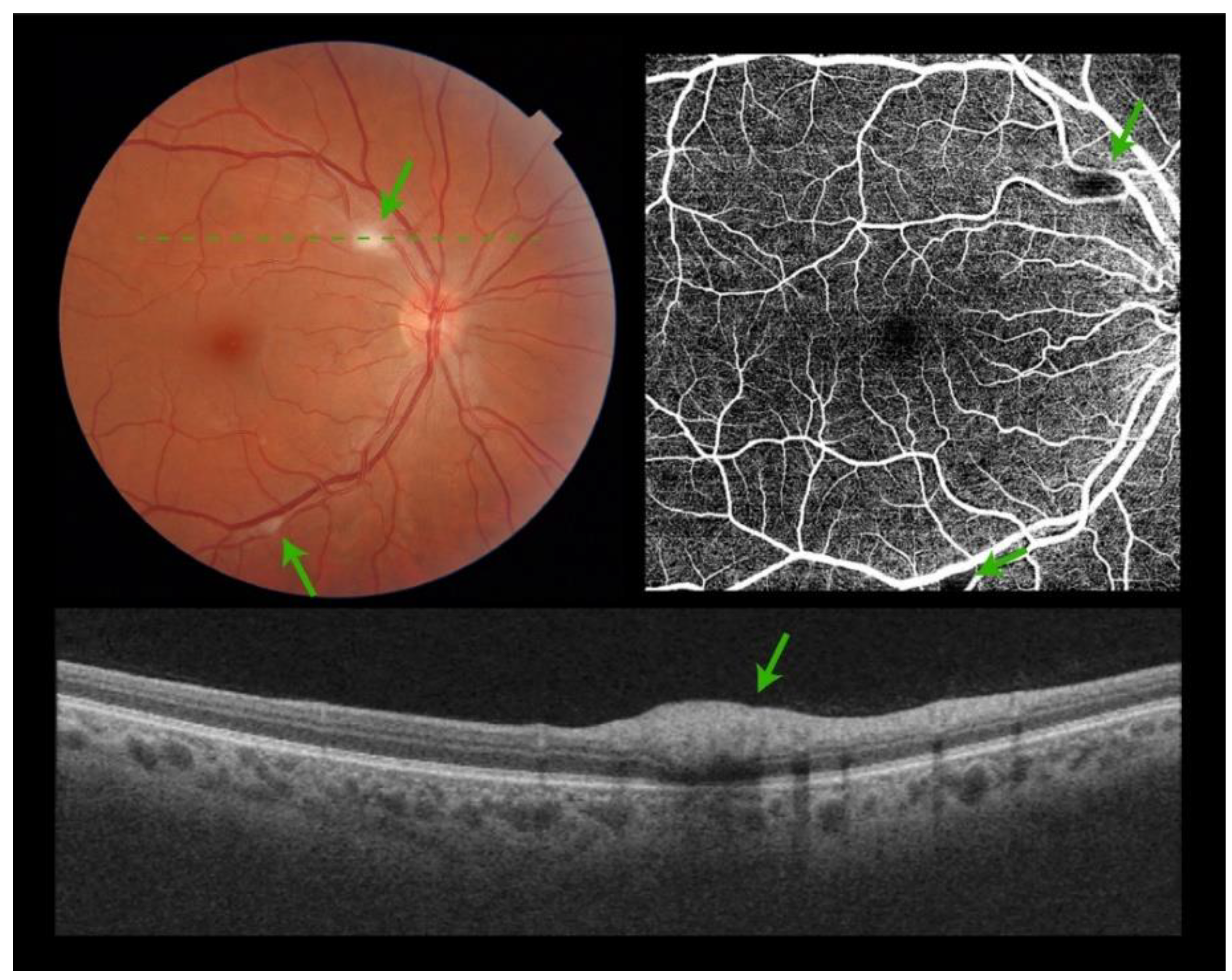
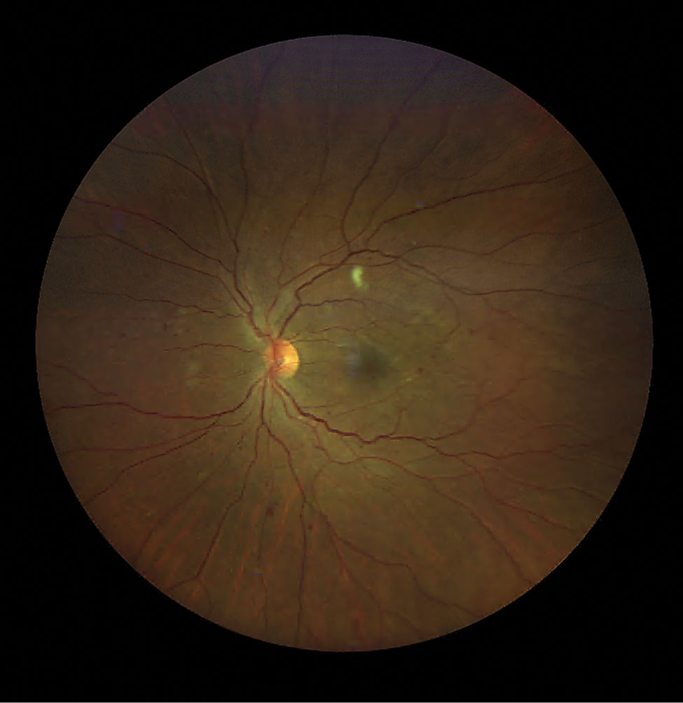
Cotton Wool Spots in a Patient with COVID-19 | Published in CRO (Clinical &  Refractive Optometry) Journal
Cotton Wool Spots in a Patient with COVID-19 | Published in CRO (Clinical & Refractive Optometry) Journal

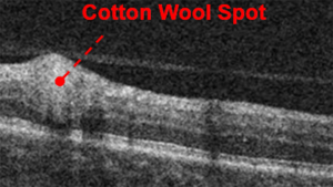
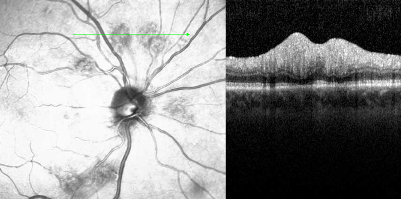
Exudates - Optical Coherence Tomography Scans

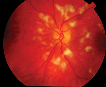
Cotton wool spots - Wikipedia

Cotton Wool Spots - EyeWiki

Right eye of a 45-year-old woman showing cotton wool spots (white... |  Download Scientific Diagram
Right eye of a 45-year-old woman showing cotton wool spots (white... | Download Scientific Diagram

Optical coherence tomography findings of bilateral foveal leukemic inf |  IMCRJ
Optical coherence tomography findings of bilateral foveal leukemic inf | IMCRJ

OCT and Me: A Beginner's Guide - mivision

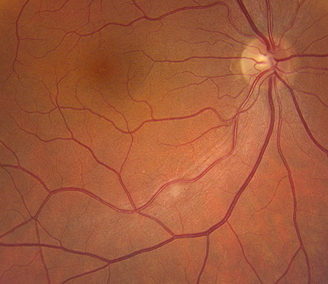
Isolated cotton-wool spot during pregnancy: A case report

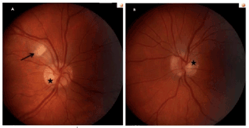
A and B) Color photograph of right and left eyes showed Purtscher-like... |  Download Scientific Diagram
A and B) Color photograph of right and left eyes showed Purtscher-like... | Download Scientific Diagram

OCT Retinal Bootcamp

Classification of Cotton Wool Spots Using Principal Components Analysis and  Support Vector Machine | Semantic Scholar
Classification of Cotton Wool Spots Using Principal Components Analysis and Support Vector Machine | Semantic Scholar

Cotton-Wool Spots - Scott E. Pautler, M.D. TampaScott E. Pautler, M.D. Tampa

Nov-2011
Nov-2011

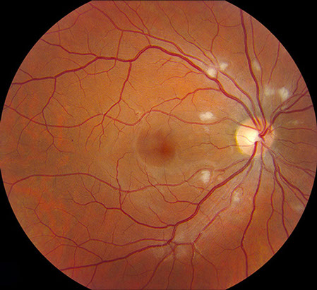
Cotton wool spots (green arrows) appear as white-green (a) or... | Download  Scientific Diagram
Cotton wool spots (green arrows) appear as white-green (a) or... | Download Scientific Diagram

A and B) Color photograph of both eyes showed Purtscher-like... | Download  Scientific Diagram
A and B) Color photograph of both eyes showed Purtscher-like... | Download Scientific Diagram

Cotton-Wool Spots May Challenge Diagnosis

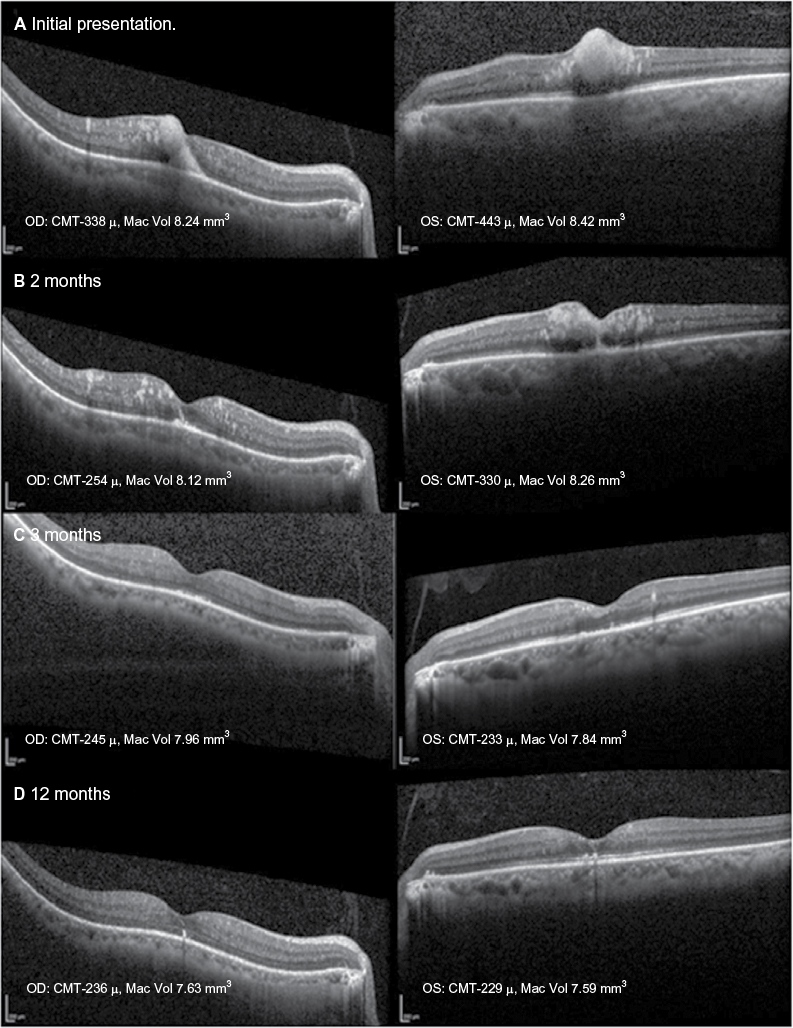
Longitudinal analysis of cotton wool spots in COVID‐19 with high‐resolution  spectral domain optical coherence tomography and optical coherence  tomography angiography - Markan - 2021 - Clinical & Experimental  Ophthalmology - Wiley Online Library
Longitudinal analysis of cotton wool spots in COVID‐19 with high‐resolution spectral domain optical coherence tomography and optical coherence tomography angiography - Markan - 2021 - Clinical & Experimental Ophthalmology - Wiley Online Library

PDF] Single Cotton Wool Spot as a Late Manifestation of Head Trauma |  Semantic Scholar
PDF] Single Cotton Wool Spot as a Late Manifestation of Head Trauma | Semantic Scholar

JCM | Free Full-Text | Retinal Manifestations in Patients with COVID-19: A  Prospective Cohort Study
JCM | Free Full-Text | Retinal Manifestations in Patients with COVID-19: A Prospective Cohort Study

Matt Hirabayashi, MD on X: "A Cotton Wool Spot occurs when changes of  retinal vasculature cause axoplasmic stasis of the RNFL. The axons swell  and cause the characteristic white spots on the
Matt Hirabayashi, MD on X: "A Cotton Wool Spot occurs when changes of retinal vasculature cause axoplasmic stasis of the RNFL. The axons swell and cause the characteristic white spots on the

Cotton-wool spots in giant cell arteritis | CMAJ

Isolated cotton-wool spot during pregnancy: A case report

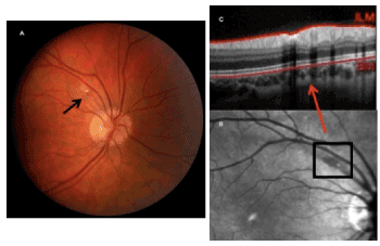
Right eye cotton-wool spot appearance on infrared imaging (A, arrow),... |  Download Scientific Diagram
Right eye cotton-wool spot appearance on infrared imaging (A, arrow),... | Download Scientific Diagram

August-2019
August-2019

Wild and Woolly
Wild and Woolly